


Examples of Stage 1 through Stage 4 dental disease
*Pets with stage 1 or 2 dental disease should be evaluated and a thorough cleaning performed prior to it reaching stage 3 (or 4) disease – Once stage 3 is reached there is irreversible damage to the tooth or bone structures which often requires tooth extraction. Routine cleanings (prior to late stage disease) and evaluation will reduce the amount of dental disease your pet experiences throughout their life, thus providing them with a healthier mouth and body.
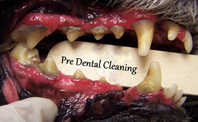
Stage 1 Dental Disease – Before Cleaning
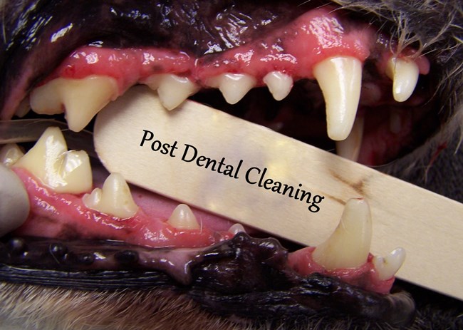
Stage 1 Dental Disease – After Cleaning – no permanent disease
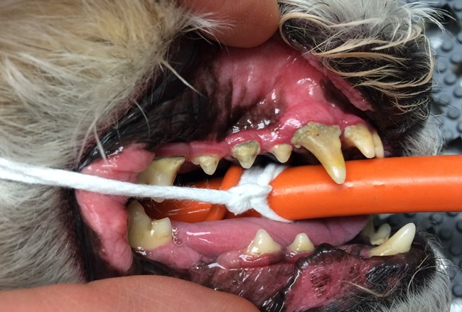
Stage 2 Dental Disease – Before Cleaning
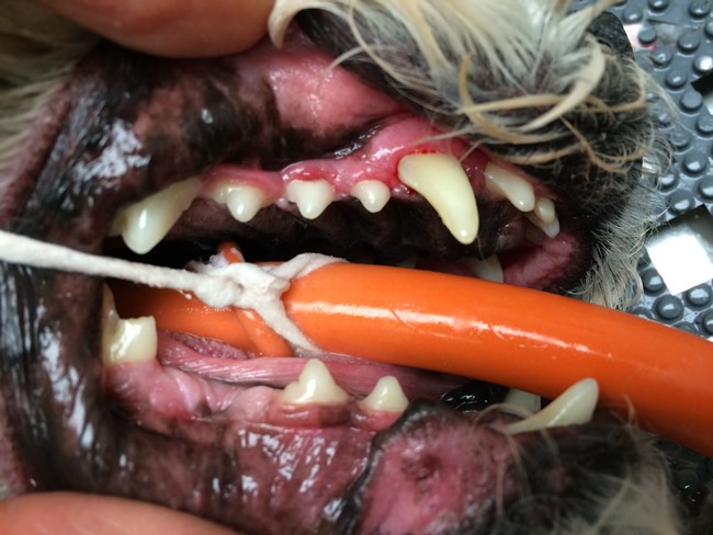
Stage 2 Dental Disease – After Cleaning – No permanent disease issues
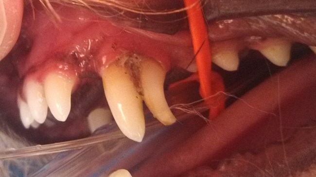
Stage 3 Dental Disease – Before cleaning – Note severe tartar and permanent gum recession.
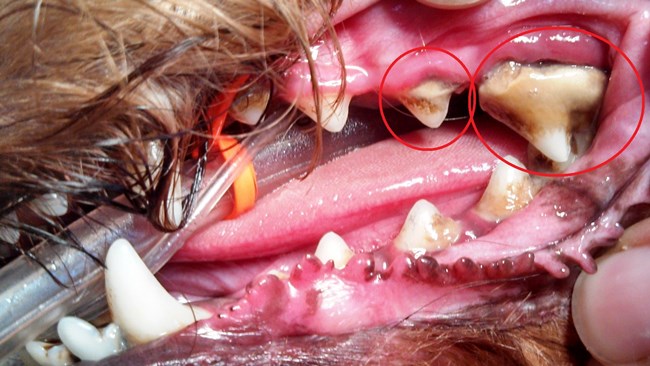
Stage 3 Dental Disease – After cleaning – The gum recession is severe, with root exposure – this tooth has permanent damage from disease and needs extracted
Stage 4 Dental Disease –
Info about above stage 4: Severe amount of tartar/calculus present covering the majority of the teeth. Multiple teeth are missing because disease caused them to fall out over time. Note the lower right canine tooth and upper premolar that is almost entirely encapsulated in heavy debris and tartar. Note gray colored, sticky, debris present along the gumline. This mouth is heavily infected and painful.
Specific examples below of different oral diseases.
Severe Periodontal Disease – After a thorough cleaning, this young dog with severe gum recession exposing roughly 80% of the length of the tooth roots. Looking in his mouth you should normally only see about 25% of the tooth length you can see here. This was all caused by gingivitis and decay from tartar and bacteria build-up. This condition results in loose teeth, pain when eating, and gingival infection. Teeth this heavily affected most commonly require extraction.
Before – note all the gray/black material between the teeth, and After – all the debris cleared away
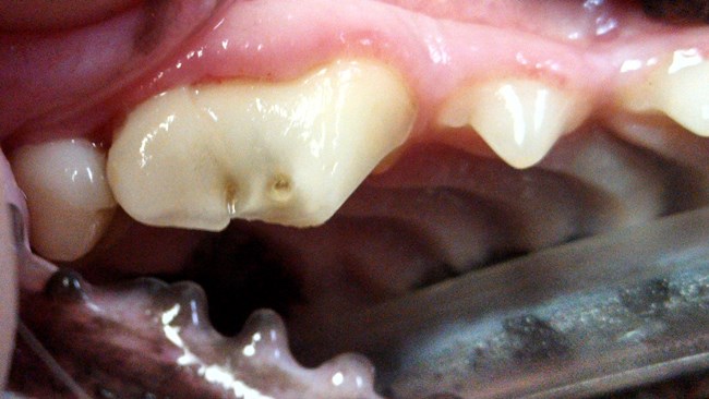
Hidden Tooth Decay – At the apex of the canine tooth you can see a dark area surrounding it – this signifies bone loss. In this patient he had chronic nasal discharge coming from this tooth root infection. This condition is painful and the jaw bone is infected. This tooth needs surgically extracted.
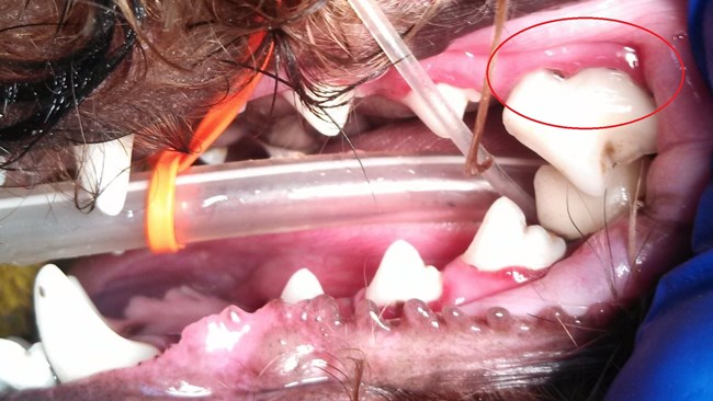
Gum Recession – Moderate gum recession (normal gum line noted by dashed line) on the canine teeth of this pet – all of the other teeth look healthy. This is a perfect example that moderate to severe disease doesn’t have to affect the whole mouth. This tooth may be able to be saved depending on several factors, or may need removed.
Loose Teeth/ Bone Loss- This lower molar in a dog is loose to the touch due to severe bone loss around the roots. This is caused by tartar and bacteria build-up that slowly destroys bone. This tooth is painful and the socket is infected.
Tooth Resorption – These teeth are resorbing from the deep aspect to the surface. This disease process takes several months to happen and is very uncomfortable during this process. The image on the left shows 2 (mostly) normal teeth, with a “bump” of bone where a tooth was previously resorbed and the material left behind is bone. The image on the right shows end stages of resorption where 90% of the tooth has been destroyed, and only the crown of the tooth remains attached to red, ulcerated, painful gum tissue. These teeth need removed to eliminate pain and infection for the pet.
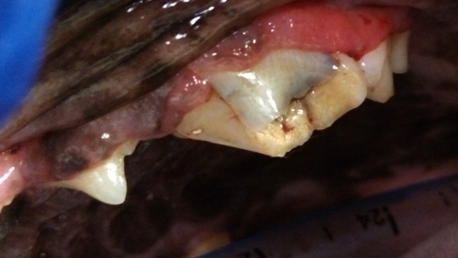
Slab Fracture – Very common fracture of the upper 4th premolar – usually as a result of chewing on objects that are way too hard for a dogs teeth. These teeth are painful, and with an exposed pulp cavity they are prone to infection.
Few Examples
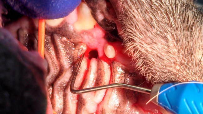
Surgery Before and After – tooth surgically removed and the ginigival flap sutured closed
Retained deciduous (juvenile) teeth –
Oral Tumor– This pet presented for an oral tumor noticed by the owner. This tumor happened to be benign (non-aggressive), however a lot of oral tumors are commonly aggressive. Many oral tumors are very difficult to remove given their location, and the fact that many of them aren’t noticed until they are relatively very large.
Misaligned teeth – This pet has severely misaligned teeth in the front part of their mouth. These incisors are not only abnormally placed and positioned, they also are trapping hair, food and debris between the tooth and the gingiva. This trapped debris causes inflammation which ultimately results in gum recession, bone loss, and loose teeth. These teeth likely need removed to restore healthy tissue to this area.
Oronasal Fistula –
Im wondering being though i had 4 pinched nerves to my left neck.Thevnew damage s on the left side of my body..Neck down arms now after bite, my nervs feel very irritated.I can feel the buttocks area pulled muscle.And the women put her hand near my leg to show the dog were to rip and lock on my leg 3 times untill he pulled the meat.I was cuffed and my dress shows her foot print pushing on my right. I want to no my energy is tired sluggish before this so i feel the bite has me in red zone.Im not safe yet